Fig. 3 .

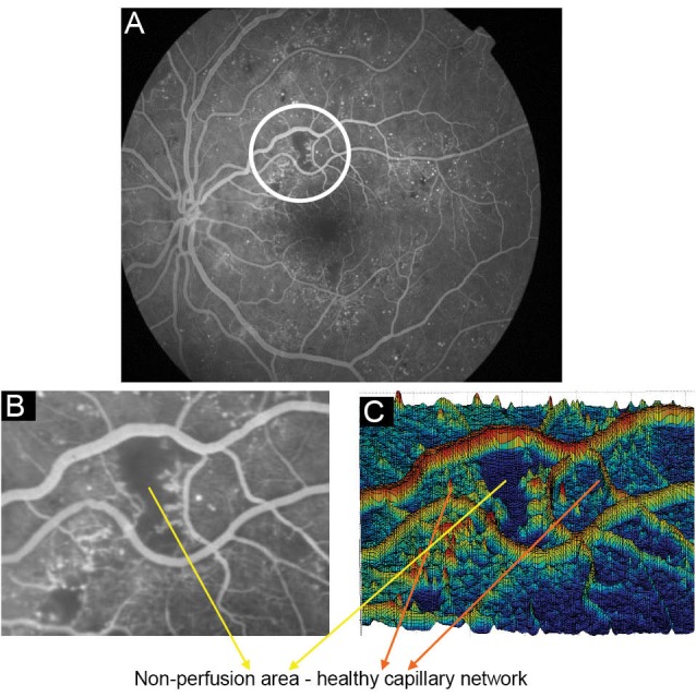
(A) A Sample FFA image with non-perfused regions, (B) A crop of the image from non-perfused area, (C) Surface of 3-D shape of non-perfusion region in the image.

(A) A Sample FFA image with non-perfused regions, (B) A crop of the image from non-perfused area, (C) Surface of 3-D shape of non-perfusion region in the image.