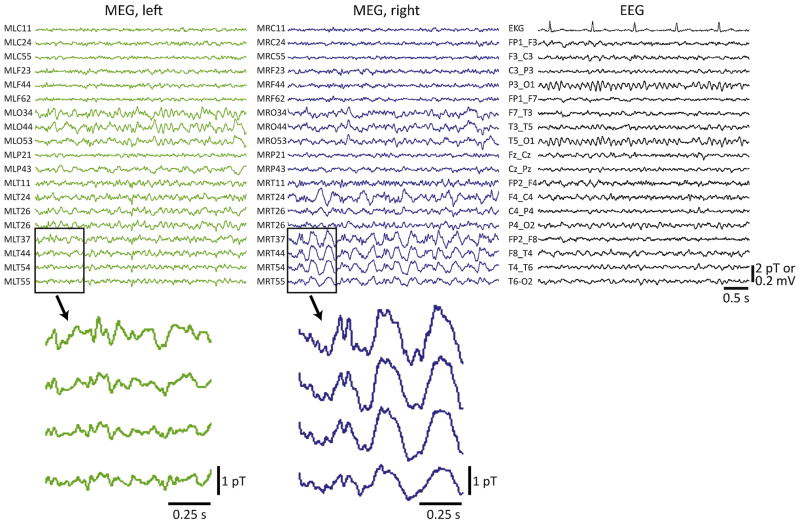
Figure 1.
Example simultaneous MEG/EEG recordings in a patient with asymmetric MEG slowing. Recordings were performed in a 33-year-old male with a seven-year history of drug resistant epilepsy and a normal MRI. MEG revealed large-amplitude slow (delta) activity in the right temporal lobe that was not appreciated on simultaneous EEG recordings (shown on the right), or on long-term scalp EEG recordings (not shown). Enlarged 1-s epochs showing a few temporal lobe channels that do not (bottom left) or do (bottom right) exhibit slow activity are also provided. No interictal spikes were captured during the MEG session. After a long-term intracranial monitoring study, the patient underwent tailored right temporal lobe resection, and remained seizure free (Engel class I outcome) four years after surgery.

