Fig. 1.

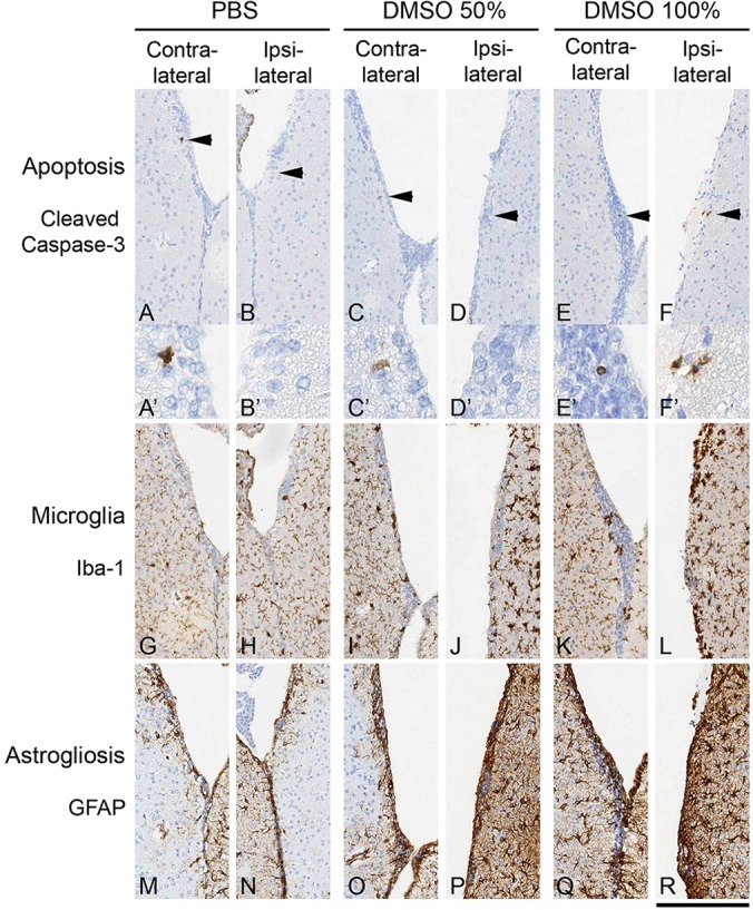
Cellular reaction to intraventricular DMSO injection. The cerebral ventricles were injected with 5 µl PBS (A,B,G,H,M,N), 50% DMSO in PBS (C,D,I,J,O,P) or 100% DMSO (E,F,K,L,Q,R). (A-F) Immunohistochemical staining for cleaved caspase 3 to detect apoptotic cells/nuclei shows occasional positive cells in controls and both DMSO groups. Arrowheads point to single positive cells in the SVZ, shown at higher magnification in A'-F'. Single cells are detected bilaterally in the SVZ. The PBS group shows a single cell contralateral to the injection (A), and there is a single cell in the contralateral wall in the 50% DMSO-injected brain, and a single cell on the contralateral side and two cells on the ipsilateral side of the 100% DMSO-injected brain. (G-L) Mice injected with DMSO show a mild microglial reaction on the injected side (J,L). (M-R) This is also associated with a mild to moderate astrocytic gliosis, which is present on the side of injection (P,R). Scale bar: 25 µm for A′-F′, and 100 µm in all other panels.
