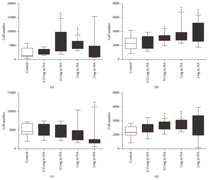
Figure 3.
MSCs grown in HA media and seeded HA media. MSCs expanded until passage 2 in control media. Cells were then stripped and cell suspensions were counted using the standard enumeration technique. Cells grown control media, flask (A), were seeded in control media (control) (− −) and MSCs grown in flasks (B–E) HA media were seeded into 96-well plates in HA media with the same concentration of HA (+ +) ranging from 0.25 to 2 mg/mL of HA to make up the five conditions (x-axis). Conditions were tested using five technical replicates per 96-well plate and run in biological triplicate (n = 3). Wells were assayed at the endpoint using Cell Counting Kit-8 with standard curves run on every 96-well plate seeded 24 hours prior to endpoint and absorbance read at a wavelength of 450 nm (y-axis). All conditions were compared back to the control using a t-test (∗ p value < 0.05, ∗∗ p value < 0.01, and ∗∗∗ p value < 0.001). (a) MSCs seeded in standard 96-well plates for 24 hours (adherence), (b) MSCs seeded in standard 96-well plates for three days (proliferation), (c) MSCs seeded in high-adherence 96-well plates for 24 hours (adherence), and (d) MSCs seeded in high-adherence 96-well plates for three days (proliferation) (see Supplementary Table 1).

