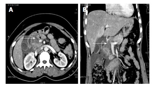
Figure 2.

A 27-year-old woman with road traffic accident. CECT axial (A) and coronal (B) images show full thickness laceration in the head of pancreas (white arrows). The laceration is located to the right of splenoportal confluence suggestive of grade IV injury. There is also fluid around the superior mesenteric vein (black arrow) and adjacent superior mesentery artery; the so-called “SMV cuff” sign seen in proximal injuries of pancreas. CECT: Contrast enhanced computed tomography; SMV: Superior mesenteric vein.
