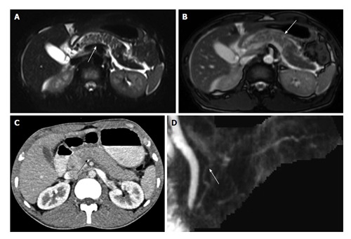
Figure 9.

A 25-year-old man was involved in road traffic accident. CECT axial image (A) shows ill-defined hypoattenuating area in neck of pancreas suggestive of contusion (arrow). MRI done 10 h after CT shows extent of contusion better with wider area of involvement on both T2 HASTE (B) and TRUFISP (C) images. MRCP thick MPR (D) image shows ductal integrity. Patient was conservatively managed and follow-up imaging showed decrease in area of contusion. CECT: Contrast enhanced computed tomography; MPR: Multiplanar reconstruction; TRUFISP: True fast imaging with steady-state free precession; MRI: Magnetic resonance imaging; MRCP: Magnetic resonance pancreatography; CT: Computed tomography.
