Figure 5.
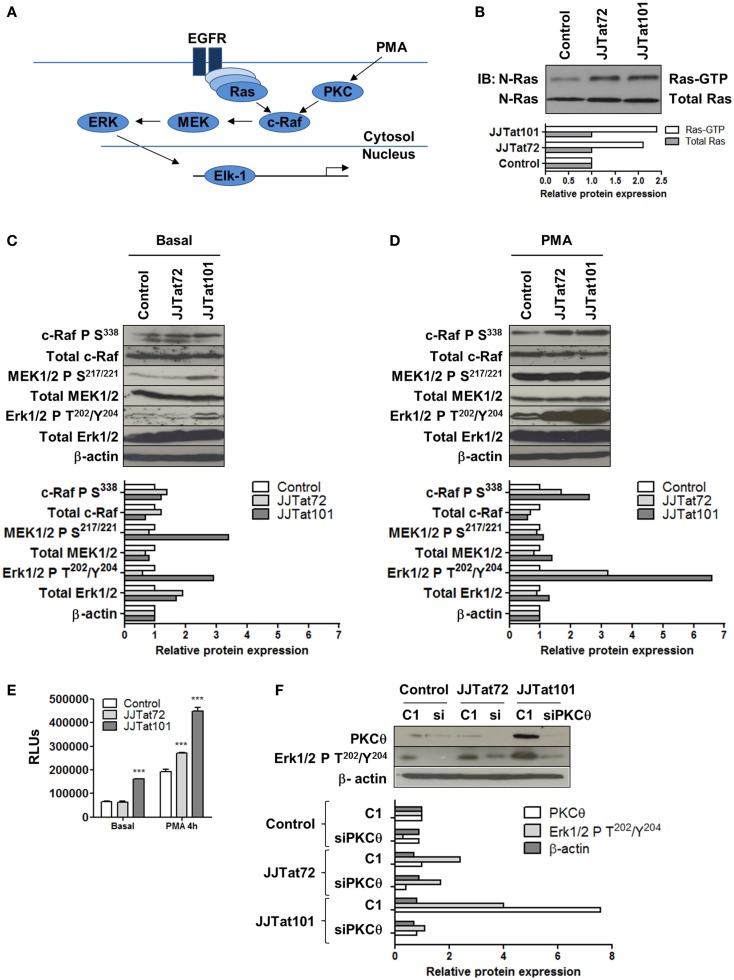
Increased activation of Ras/Raf/MEK/ERK/Elk-1 pathway in Jurkat-Tat cells was dependent on PKCθ activation. Schematic representation of the Ras/Raf/MEK/ERK/Elk-1 pathway (EGFR, epidermal growth factor receptor) (A). Analysis by immunoblotting of Ras-GTP activity in Jurkat-Tat cells (B). Analysis by immunoblotting of phosphorylated and total proteins of the Ras/Raf/MEK/ERK pathway in Jurkat-Tat cells in basal conditions (C) and after activation with PMA for 15 min (D). Analysis by chemiluminescence of Elk1 activity in Jurkat-Tat cells in basal conditions and after treatment with PMA. Data are represented as mean ± SEM. *** for p < 0.001 (E). Analysis by immunoblotting of levels of ERK1/2 phosphorylation at T202/Y204 after RNA interference of PKCθ in Jurkat-Tat cells (F). Densitometry analyses were performed to calculate relative protein expression and showed as horizontal bar diagrams (B–D,F).

