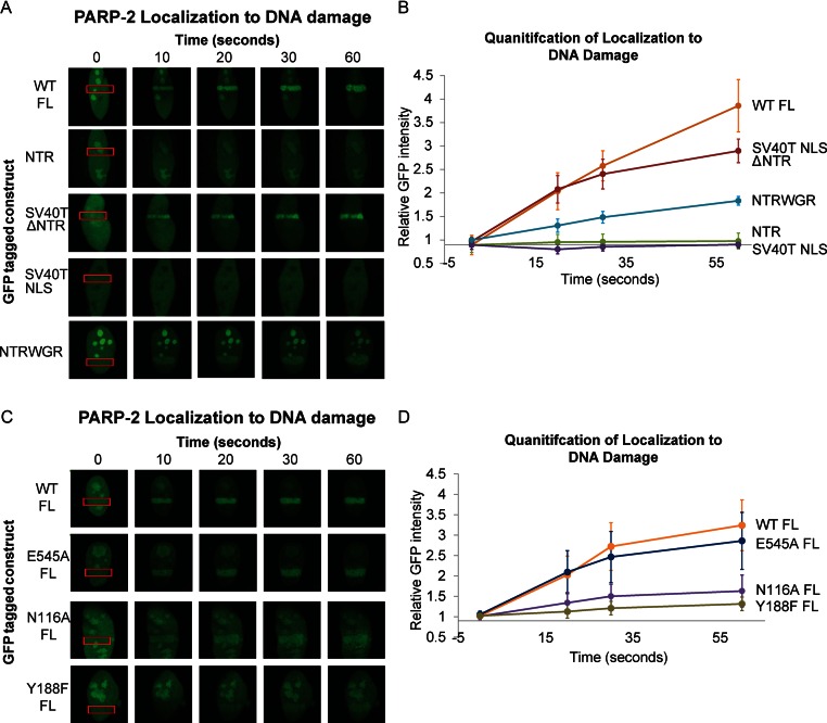
Figure 4.
PARP-2 recruitment to cellular sites of DNA damage. (A) and (C) Live-cell imaging of recruitment of GFP-PARP-2, truncation constructs (A) or FL mutants (C) to sites of laser-induced DNA damage. The region of laser irradiation is indicated with a red box. The images were captured at the various time points indicated. The images shown are representative the results obtained from at least three independent experiments. SV40T NLS is an N-terminal peptide tethered to PARP-2 truncations that lack the canonical NLS of PARP-2. (B) and (D) Quantitation of relative GFP intensity within the laser path relative to background (a non-irradiated area of the nucleus). Relative GFP signal averaged for ≥4 cells. Error bars represent the SD.

