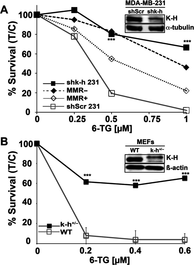
Figure 2.

K-H knockdown cells exhibit DNA damage tolerance following 6-TG exposure. (A) shScr or shk-h knockdown 231 TNBC cells and MMR-deficient or MMR-proficient colon cancer cells (as controls) were seeded onto 48-well plates. 6-TG (μM) was added ∼12 h later and cells were exposed for 48 h. After drug removal, cells were grown for 7 days. Relative survival, measured by DNA content, was monitored using Hoechst 33258 dye staining and a Victor X3 plate reader (48). Data were expressed as means, ±SD for treated/control (T/C) cells from experiments performed at least 3 times in triplicate. P-values were obtained via two-tailed Student's t-tests. ***P < 0.001, comparing shScr versus shk-h. K-H and α-tubulin (loading) levels were monitored to compare K-H expression knockdown in shScr versus shk-h cells. (B) Mouse embryonic fibroblasts (MEFs) derived from WT (mk-h+/+) or mk-h+/− heterozygous mice (8) were used to measure sensitivities to 6-TG exposures. Relative cell survival assays were then performed as in Figure 2A. K-H protein levels in MEFs were analyzed by Western blotting using an antibody against human K-H protein. β-Actin was used for loading. K-H levels in these cells were previously confirmed using specific antibodies (8). Antibodies against human K-H also work against mouse K-H due to the conserved nature of the protein (8).
