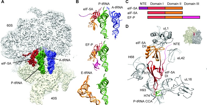
Figure 1.
Cryo-EM structure of eIF-5A bound to the yeast 80S ribosome. (A) Transverse section of the cryo-EM map of eIF-5A–80S complex, (40S, yellow; 60S, gray), revealing the binding site of eIF-5A (dark red), P-tRNA (green) and A-tRNA (blue). (B) Comparison of ribosome binding positions of eIF-5A, EF-P (16) and E-site tRNA(48), relative to A-tRNA (blue) and P-tRNA (green). The domains for eIF-5A and EF-P are colored according to (C). (C) Schematic representation of the domain structures of eIF-5A, aIF-5A and EF-P. (D) Molecular model for the interaction of domains I (DI, red) and II (DII, orange) as well as N-terminal extension (NTE, magenta) of eIF-5A with rRNA and ribosomal protein components of the ribosome (gray). Ribosomal insert shows the orientation of the view.

