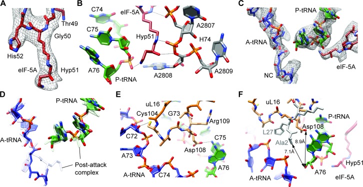
Figure 3.
Hypusine of eIF-5A at the peptidyltransferase center of the ribosome. (A) Electron density (gray mesh) and molecular model (red) for hypusine 51 (Hyp51) of eIF-5A. (B) Potential hydrogen bond interactions (dashed lines) between hypusine 51 (Hyp51) of eIF-5A (red) with the CCA-end of the P-tRNA (green) and 25S rRNA nucleotides within helix H74 (gray). (C) Electron density (gray mesh) and molecular models for the CCA-ends of P-tRNA (green) and A-tRNA with nascent chain (NC) (blue) as well as eIF-5A (red). (D) Comparison of A-tRNA (blue) and P-tRNA (green) from eIF-5A–80S complex with A- and P-tRNAs from post-attack complex (33) (gray). (E) Potential hydrogen bond interactions (dashed lines) between the loop of uL16 (orange), A-tRNA (blue) and P-tRNA (green). (F) Proximity of Asp108 of uL16 (orange) to the 3′ OH of A76 of the P-tRNA (8.9 Å) in the eIF-5A–80S complex, compared with the proximity of Ala2 of L27 (gray) to the 3′ OH of A76 of the P-tRNA (7.1 Å) in the pre-attack complex (33). The CCA-end of the A-tRNA (blue) and hypusine 51 (Hyp51) of eIF-5A (red) are shown for reference.

