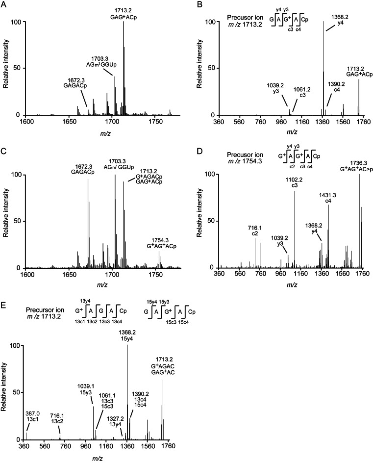
Figure 8.
MALDI-MS analysis of the wild-type tRNALeu. (A) The wild-type tRNALeu was expressed in T. kodakarensis strain KUWA and purified as a control. The purified tRNALeu was digested with RNaseA and its fragments were then analyzed by MALDI-MS spectrometry. In this region, a fragment (m/z = 1713.2), which coincided with the expected m/z value of the G13-C17 fragment (GAG+ACp) in the wild-type tRNALeu, was detected. (B) The fragment sequence (m/z = 1713.2) was determined by MS/MS analysis as GAG+ACp. (C) The RNaseA-digested fragments derived from the wild-type tRNALeu expressed in the KTA1493 strain were analyzed by the same method as (A). In the 1600–1800 m/z region, a new fragment (m/z = 1754.3) appeared. (D) The sequence of this fragment (m/z = 1754.3) was determined as G+AG+ACp, which corresponded to the G+13-C17 fragment in the wild-type tRNALeu. (E) Two fragments (G+AGACp and GAG+ACp; m/z = 1713.2) were detected by MS/MS analysis.

