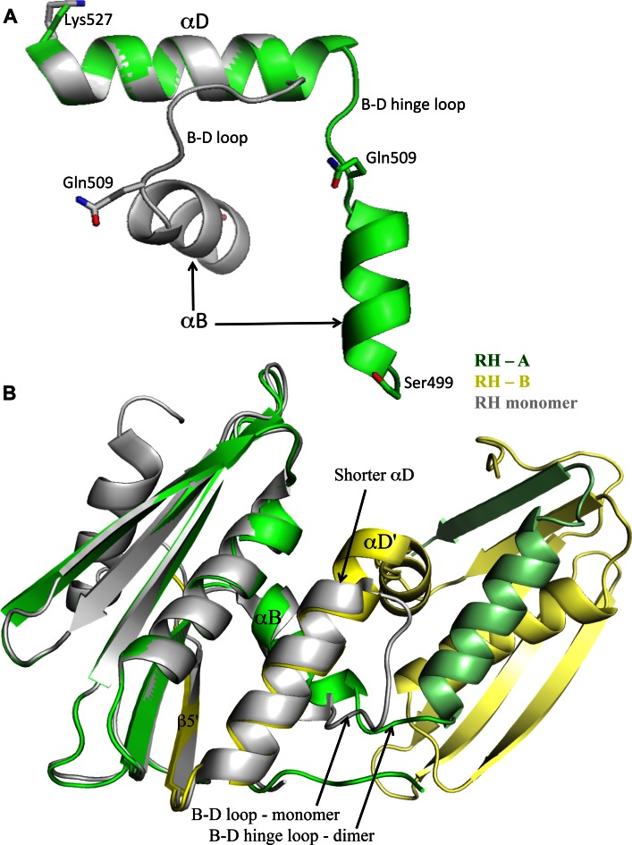
Figure 4.
Shortening of the B-D hinge loop. (A) Ribbon representations of the αB-αD regions of one molecule of the dimer (green) and the RH monomer (gray) in which the αD helices have been aligned. As is apparent from the figure, several residues that are part of the B-D loop in the monomer are incorporated into an extended αD in the dimer. The reduction in length of the dimer B-D linker is also apparent in Figure 2B. (B) Ribbon diagram overlay of the RH monomer (gray, pdb: 3K2P) with the domain swapped dimer illustrating the change in length of αD and the B-D linker.

