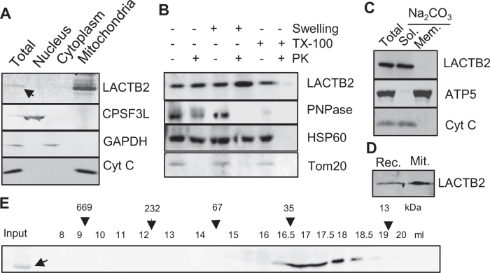
Figure 1.
LACTB2 is a mitochondrial, soluble and monomeric protein. (A) Mitochondria localization. HeLa cells were disrupted and the nuclear, cytoplasmic and mitochondria-containing fractions were separated and analyzed for the localization of LACTB2, using an immunoblotting assay. The nucleus-located protein, CPSF3L, was used as marker for nuclear proteins. Glyceraldehyde-3-phosphate dehydrogenase (GAPDH) served as a cytosolic marker and cytochrome C (Cyt C) as a mitochondrial marker. (B) Immunoblot analysis of mitochondrial extract following Proteinase K accessibility assay. TX-100, Triton X-100. PK, Proteinase K. PNPase is a mitochondrial protein located mainly in the intermembrane space and a small amount in the matrix. HSP60 is a mitochondrial matrix protein. Tom20 is a mitochondrial outer membrane protein. (C) LACTB2 is a soluble protein. Immunoblotting analysis of mitochondrial extract following alkaline sodium carbonate (Na2CO3) extraction of soluble and peripheral membrane proteins. T, total extract. S, supernatant. P, pellet. Subunit 5 of the ATP synthase (ATP5) was used as a marker for intrinsic membrane proteins and Cyt C as a marker of soluble proteins. (D) Immunoblot analysis of the recombinant LACTB2 (Rec.) and the native protein in isolated mitochondria (Mit.) showing the same size on SDS-PAGE. Therefore, the mitochondria targeting signal is not cleaved upon entering the mitochondria and remain an integral part of the mature protein. (E) LACTB2 is present as a monomer. Soluble proteins of bovine mitochondrial matrix were extracted from bovine liver as described in the methods section. This extract was fractionated on a size exclusion column superdex 200 and analyzed for LACTB2, using an immunoblotting assay. The following proteins were used as size markers: Tyroglobulin (669 kDa), polynucleotide phosphorylase (PNPase) (232 kDa), BSA (67 kDa), β-lactoglobulin (35 kDa) and ribonuclease A (13 kDa).

