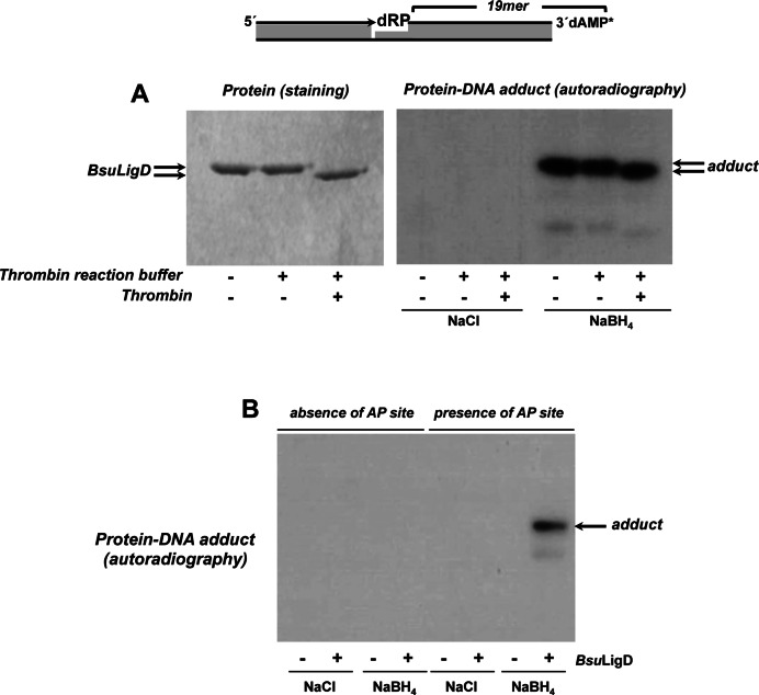
Figure 3.
Formation of BsuLigD-DNA adducts. (A) Dependence of BsuLigD-DNA cross-link on NaBH4. Reactions were performed as described in Materials and Methods, incubating 95 nM BsuLigD with 2.6 nM of the 3′ [α32P]3′-dAMP labeled DNA substrate depicted on top of the figure, in the presence of 10 μM CTP, 0.64 mM MnCl2 and either 100 mM NaBH4 or NaCl (as indicated). Left panel: Coomassie blue staining after SDS–PAGE of purified BsuLigD. Right panel: autoradiography of corresponding protein-DNA adducts after the SDS–PAGE separation shown in left panel. When indicated, protein was previously incubated with 0.05 U of thrombin at 20°C for 60 min. (B) Adduct formation is dependent on the presence of an abasic site. Reactions were performed as in described in (A) but using as substrate 3.6 nM of the 3′ [α32P]3′-dAMP labeled oligonucleotide without removing the uracil (absence of AP site) or after treatment with E. coli UDG (presence of AP site), in the presence of either 100 mM NaBH4 or NaCl (as indicated). Autoradiography of corresponding protein-DNA adduct after the SDS–PAGE separation is shown.

