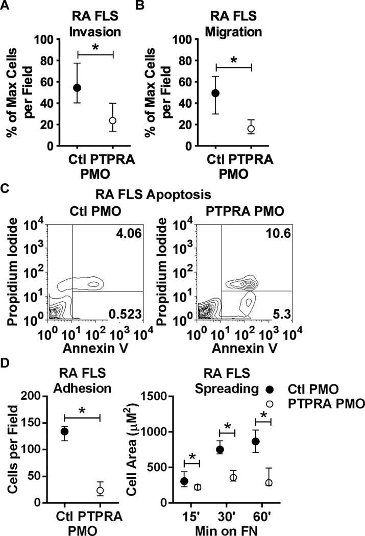
Figure 2. RPTPα promotes RA FLS invasiveness.
(A) Following treatment with PMO for 7 d, RA FLS (n=4) invaded through Matrigel-coated transwell chambers in response to 50 ng/ml PDGF-BB for 48 hr. (B) PMO-treated RA FLS (n=4) migrated through uncoated transwell chambers in response to 5% FBS for 24 hr. (A–B) Median and IQR % maximum number of cells per field is shown. *, p<0.05, Mann-Whitney test. (C) PMO-treated RA FLS were washed and stimulated with 50 ng/ml PDGF for 24 hr. Cells were collected and stained with Annexin V and PI, and cell fluorescence was assessed by FACS. Graphs show gating strategy to detect early apoptotic (Annexin V+PI−) and necrotic/late apoptotic (Annexin V+PI+) cells. Significance was calculated using the Chi square test (p<0.0001, Chi-square=2294, df=2). Data is representative of 4 independent experiments. (D) PMO-treated RA FLS (n=4) were plated on fibronectin (FN)-coated coverslips in the presence of 5% FBS. Graphs show median and IQR cells per field after 15 min (left) or cell area after 15, 30 and 60 min (right). *, p<0.05, Wilcoxon matched-pairs signed rank test.

