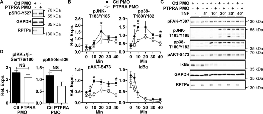
Figure 3. RPTPα promotes RA FLS signaling downstream SRC.
(A) anti-pSRC-Y527 levels in PMO-treated RA FLS lysates were measured by Western blotting. Data is representative of 4 independent experiments. (B–C) Western blotting of lysates of PMO-treated RA FLS stimulated with 50 ng/ml TNF or left unstimulated. (B) Signal intensities of Western blots of TNF-activated proteins from lysates were quantified by densitometric scanning. Mean ± SEM of signal relative to GAPDH from 6 RA FLS lines is shown. (C) Representative image is shown. (D) Signal intensities of Western blots of lysates from unstimulated PMO-treated RA FLS. Mean ± SEM of signal relative to p65 from 6 RA FLS lines is shown. *, p<0.05; NS, non-significant, Wilcoxon matched-pairs signed rank test.

