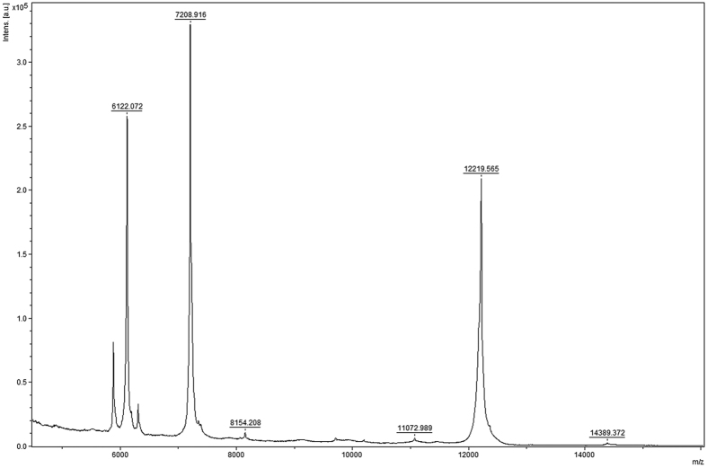
Figure 11. An example of an experimental mass spectrum of a protein after proteolytic cleavage.
Ribonuclease binase from B.pumilus was cleaved by trypsin and the cleavage fragments were analysed on HPLC LC-MS/MS system (Bruker, Germany)60. The numbers denote determined oligopeptide masses.

