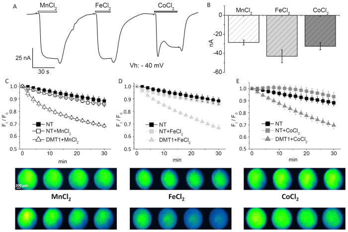
Figure 1. Calcein as a cytoplasmic “metal detector”.
(A): two electrode voltage clamp of a representative rDMT1 transfected oocyte; inward currents are induced by 100 μM MnCl2, FeCl2 and CoCl2 (Vh = −40 mV, pH 5.5). (B): means and standard errors of the transport currents obtained from 40 oocytes, five batches. (C–E): Plots of fluorescence decay (Ft/F0) with corresponding images of Calcein-injected oocytes (upper series: non-transfected (NT) and lower series: rDMT1 transfected) exposed to 100 μM MnCl2 (C), FeCl2 (D), and CoCl2 (E) at pH 5.5 from 3 to 10 oocytes, from 2 to 4 oocytes batches.

