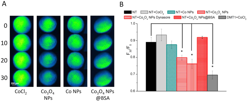
Figure 2. Calcein quenching in oocytes exposed to cobalt NPs.
(A): Representative image series of non-transfected (NT) Calcein-injected oocytes exposed to CoCl2 or different cobalt NPs for 0, 10, 20 and 30 min. (B): Means of the fluorescence decay of 5 to 25 oocytes (obtained from 2 to 5 different batches). Decay is expressed as the fluorescence intensity at time 30 min over fluorescence intensity at time 0 (F30/F0). Note that quenching is statistically significant in NT oocytes exposed to bare Co3O4 NPs and in rDMT1 expressing oocytes exposed to CoCl2 (positive control); moreover, the endocytosis blocker Dynasore does not change the quenching effect of Co3O4 NPs. Bars are ± SE; stars indicate a statistically significant (One-way ANOVA, P < 0.05) difference with non exposed oocytes.

