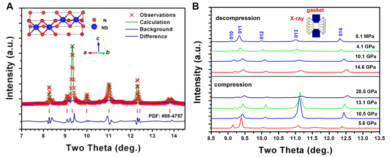
Figure 2.
(A) Refined synchrotron angle-dispersive X-ray diffraction pattern of NbN powder at ambient conditions, suggesting a hexagonal-structured ε-NbN: (Space group: P63/mmc, No. 194). Red crosses and olivine lines denote the observed and calculated profiles, respectively. The red tick marks correspond to the peak positions of hexagonal ε-NbN (PDF: #89-4757). The inset is the crystal structure of hexagonal ε-NbN. (B) Selected synchrotron angle-dispersive X-ray diffraction patterns of ε-NbN upon compression up to ∼20.5 GPa, in comparison with those during decompression where the peaks of the hexagonal phase were indexed (PDF: #89-4757).

