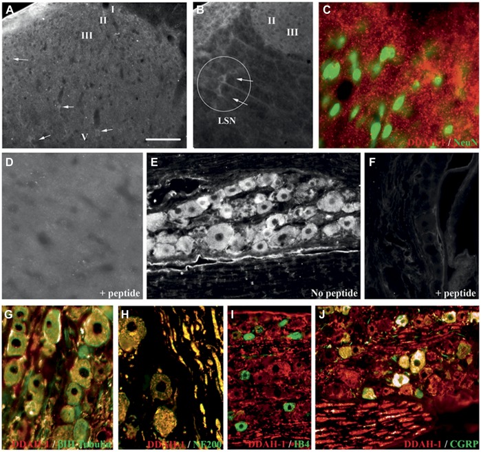
Figure 2.
DDAH-1 is expressed in sensory neurons within the DRG and dorsal horn. (A-J) Expression of DDAH-1 in the rat spinal cord and DRG. (A) Immunofluorescent staining of DDAH-1 in the lumbar spinal dorsal horn is localized in weakly stained neuronal soma in the grey matter. (B) Neurons stained in the lateral spinal nucleus (LSN) display stronger staining in the dorsal horn. (C) Merged double staining image of DDAH-1 (red) and NeuN (green, marker of neuronal nuclei) in the dorsal horn. (D, F) Specificity of the anti-DDAH-1 antibody demonstrated by preabsorption with DDAH-1 peptide and subsequent loss of staining in (D) spinal cord and (F) DRG. (E) Immunofluorescent staining of DDAH-1 in DRG neurons. (G-J) Merged double staining images of DDAH-1 (red) in DRG with (G) β-III Tubulin (green, neuronal marker); (H) with NF200 (green, marker of large myelinated DRG neurons); (I) with IB4 (green, marker of small nonpeptidergic DRG neurons); (J) with CGRP (green, marker of small peptidergic DRG neurons). Scale bar = (A): 70 μm, (B): 78 μm, (C, D): 28 μm, (E, F): 70 μm, (G): 42 μm, (H): 55 μm, (I): 97 μm, (J): 61 μm.

