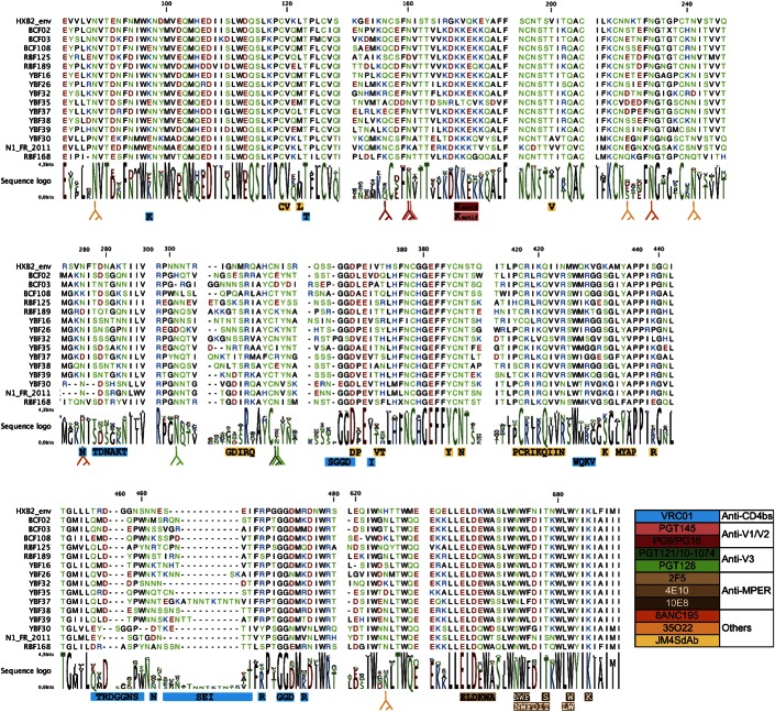
FIGURE 2.
Conservation of amino acids involved in antibody binding epitopes. An alignment of the env protein sequences of the non-M viruses used in the study is depicted, with dashes representing gaps introduced to improve the alignment. HXB2 sequence is shown as reference. Amino acids are colored based on their physicochemical properties. The logo plots denote the conservation of individual amino acids, with the height of each letter indicating the proportion of sequences that contain the residue at that site. Contact residues of VRC01 (blue), JM4SdAb (yellow), and MPER bAbs (brown) are highlighted below the alignment. The Y symbols indicate the positions of the glycans associated with antibody neutralizing activity (see colors in the inserted legend to identify the corresponding bNAbs).

