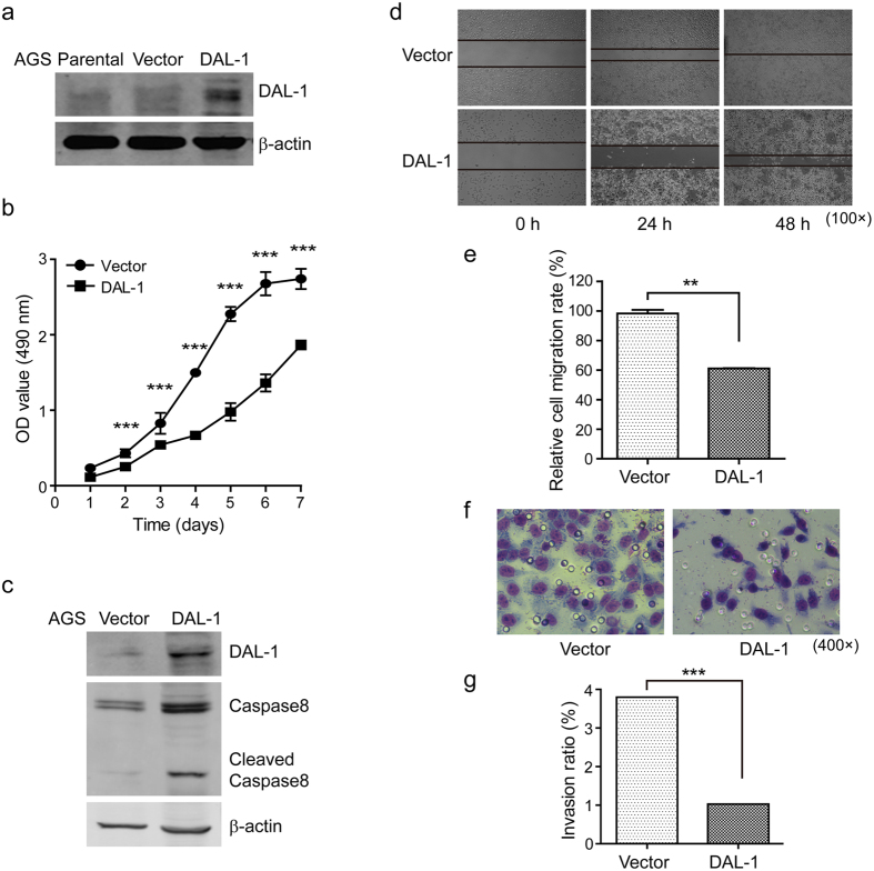
Figure 4. Overexpression of DAL-1 decreases the malignancy potential of AGS cells.
(a) DAL-1 expression in AGS cells transfected with the DAL-1 and control vector. (b) The growth rate of AGS cells. (c) The expression of caspase-8 in transfected AGS cells. (d) The migrating cells obtained at the indicated time points after wound formation. (e) The percentage of the migration rate. (f) The invading cells passing through the matrigel-coated membrane. (g) The percentage of the invasion rate. **P < 0.01, ***P < 0.001, with t-test analysis.

