
Figure 3.
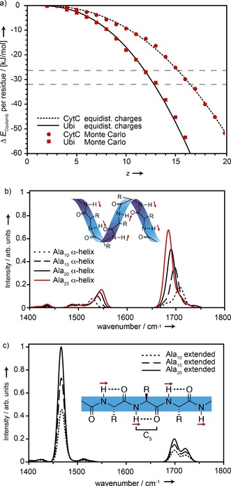
a) Difference in Coulomb energy (ΔE C) per amino acid residue between helical and extended conformations of ubiquitin and cytochrome c. The dashed horizontal lines indicate helix stabilization energies obtained from quantum chemistry calculations.10 b,c) Calculated IR spectra for neutral polyalanine in α‐helical (b) and extended (c) conformations. Shown schematically as red arrows are the directions of the amide II transition dipoles. C5 hydrogen bonds for extended structures are shown schematically as dotted lines.