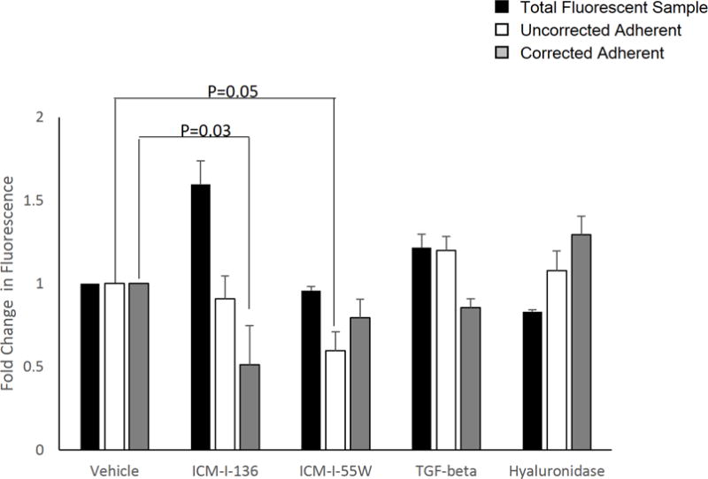
Figure 2. Soluble peptides and chemical inhibitors alter calcein fluorescence in a cell-based adhesion assay.

LNCaP cells or sublines were treated with VSSC inhibitors ICM-I-136 and ICM-I-55W, TGF-β1, hyaluronidase, or the respective vehicle, and labeled with calcein AM as described. Data were analyzed in three ways: (1) the fluorescence measurement of the treated adherent sample was normalized directly to that of the respective vehicle-treated adherent sample (uncorrected adherent, white bars), (2) the fluorescence measurement of the treated sample of 1 × 10⁵ cells was normalized to that of the respective mock or vehicle-treated sample in order to analyze the effect of the treatment on intracellular calcein fluorescence with a defined number of cells (total fluorescent sample, black bars), or (3) the fluorescence of each treated/mock-treated adherent sample was first normalized to its respective sample of 1 × 10⁵ cells (of the same treatment group) to obtain the percentage of adhered cells, and subsequently the percentage of adherent cells for the treatment group was normalized to that of the respective vehicle control to obtain fold change in adhesion (corrected adherent, gray bars). Values and error bars represent the mean fold change and standard error of three independent experiments performed in triplicate.
