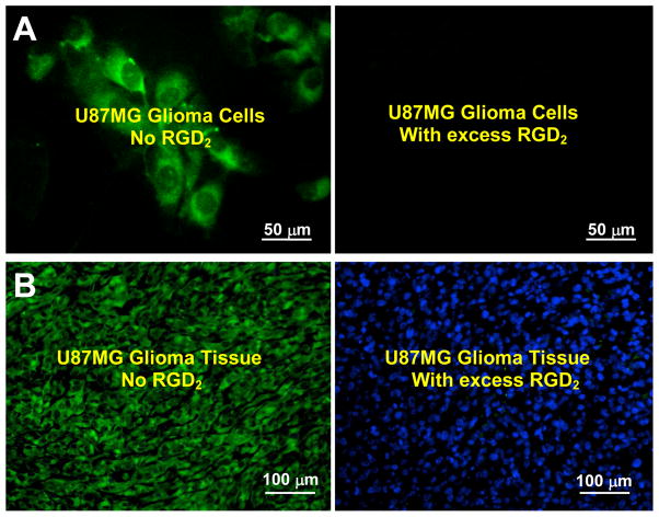
Figure 12.
A: Selected microscopic images (Magnification: 400×) of living U87MG glioma cells stained with FITC-Galacto-RGD2 in the absence (left) and presence (right) of excess RGD2. B: Microscopic images (Magnification: 200×) of a tumor slice stained with FITC-Galacto-RGD2 in the absence (left) and presence (right) of excess RGD2. The cellular and tumor staining data were from reference 183. C: Comparison of organ uptake (%ID/g) for 99mTc-2P-RGD2 in the absence or presence of excess RGD2 at 60 min p.i. D: The 60-min planar images of the tumor-bearing mice administered with 99mTc-3P-RGD2 in the absence/presence of RGD2. Co-injection of excess RGD2 resulted in significant reduction in the uptake of 99mTc-3P-RGD2 in both tumor and normal organs. The biodistribution and imaging data were obtained from reference 35.


