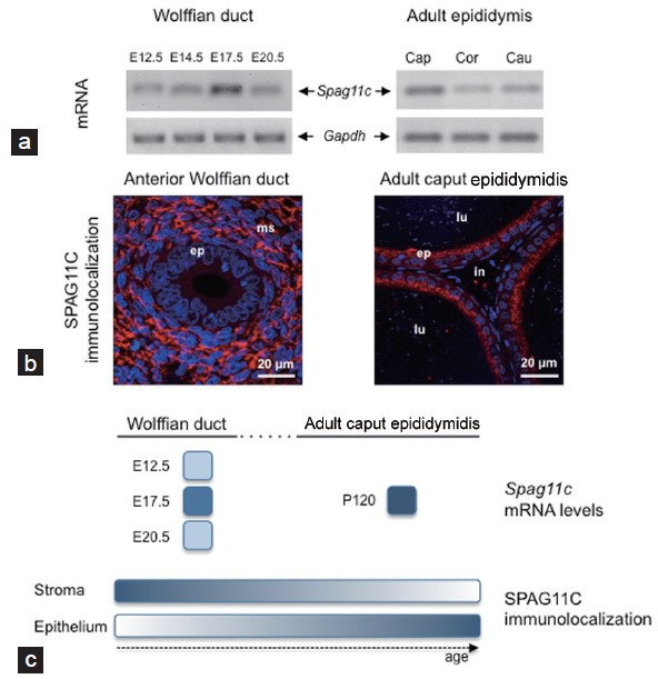
Figure 1.

Spatiotemporal expression of the β-defensin SPAG11C in the developing rat epididymis. (a) Spag11c mRNA was detected by end-point RT-PCR in the male Wistar rat urogenital rudiment as early as embryonic day 12.5 (E12.5), before the onset of androgen receptor (AR) expression and testosterone synthesis in the embryo. Increased Spag11c mRNA levels were observed in the Wolffian duct (WD) at E17.5, in contrast to decreasing levels detected at E20.5, when the WD morphologically differentiates into the epididymis under androgen influence. In the adult rat epididymis, Spag11c mRNA was more abundant in the caput (Cap) than in corpus (Cor) or cauda (Cau) epididymidis. The housekeeping gene Gapdh was used as an endogenous control. (b) Immunofluorescence studies performed in paraffin-embedded sections from whole fetuses (E18.5) and adult caput epididymidis (120 days old, P120) revealed that SPAG11C was prenatally mainly located in mesenchymal cells of the anterior WD (the future epididymis) (left panel), shifting gradually to epithelial cells after birth and became mainly distributed in the epithelium of adult caput epididymidis (right panel). Nuclei were stained with DAPI (blue). lu: lumen; ms: mesenchyme; ep: epithelium; in: interstitium. (c) Schematic representation of SPAG11C (mRNA levels and immunolocalization) developmental changes in the rat WD and adult epididymis. Relative expression levels were represented by a gradient of blue shades ranging from white (minimal intensity) to dark blue (maximal intensity) based on RT-qPCR data.44 P: postnatal day. Results from panels (a) and (b) are representative of previously published data by our group.44
