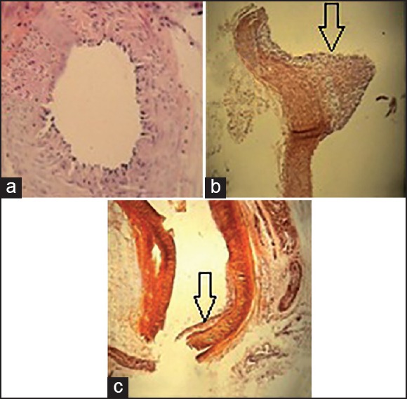
Figure 1.

Pathological evaluation of renal arteries in all groups: (a) normal control group, (×40) normal arterial wall, (b) hypercholesterolemic control group (×10) highly protruding atherosclerotic plaque, (c) treatment group (×10) foam cells in intima. Arrows: Atherosclerotic plaques
