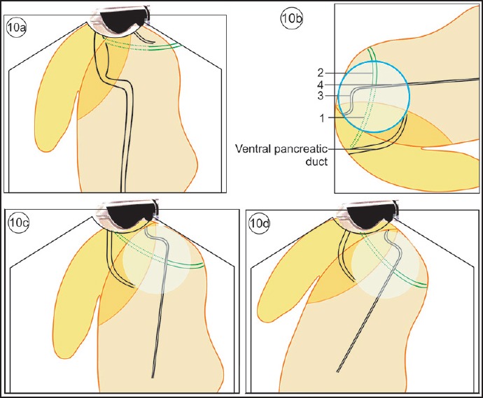
Figure 10.

(a) The VP duct lies beyond the bile duct and remains parallel to the CBD. The DP duct is smaller in diameter and tapers within the pancreatic parenchyma. (b) The circle within which the crossing over of the duct occurs is seen. This crossing over is also visualized in MRCP. The four parts related to the crossing are the intrapancreatic or retropancreatic CBD, CBD within hepatoduodenal ligament, the dorsal pancreatic duct before crossing, and the dorsal pancreatic duct after crossing. (c) The imaging of “cross duct sign” from the second part of duodenum shows the crossing over of pancreatic duct. (d) The imaging of “cross duct sign” from the duodenum bulb shows the crossing over of pancreatic duct with the bile duct
