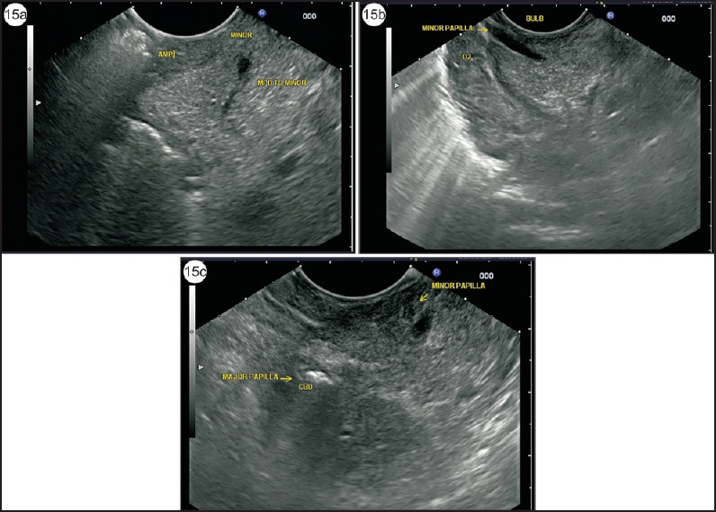
Figure 15.

(a) Application of color Doppler can identify the small anechoic structures when the scope is wedged into the duodenal bulb in a long loop position or during withdrawal from the second part of the duodenum. The duct of minor papilla usually ends in a triangular hypoechoic area near the duodenal wall. The duct is seen in a transverse axis. (b) In such cases, an anechoic duct going toward the duodenal wall within the parenchyma of hyperechoic DP is the duct of Santorini. The duct is seen on a longitudinal axis. (c) This image shows both major and minor papilla, which are placed at a distance of about 1.5 cm. The air is seen in CBD
