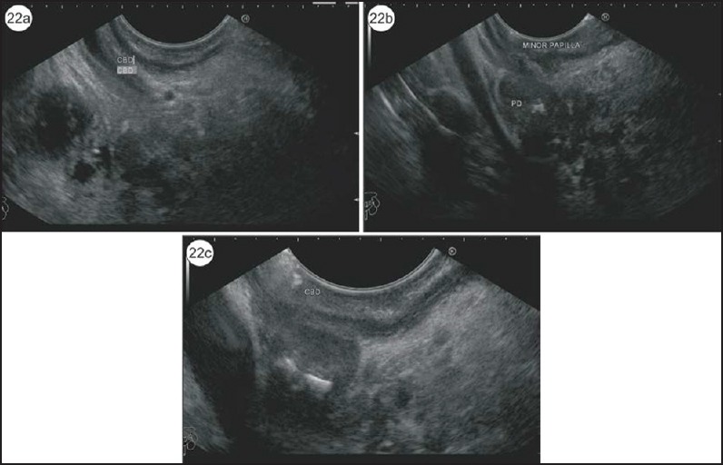
Figure 22.

(a) In this case of PD, initially the CBD is identified in the duodenal bulb. (b) The pancreatic duct with calculi is identified in the same position a little cranial to the CBD. (c) This is a case of PD with santorinicele and a stone lying just above the santorinicele
