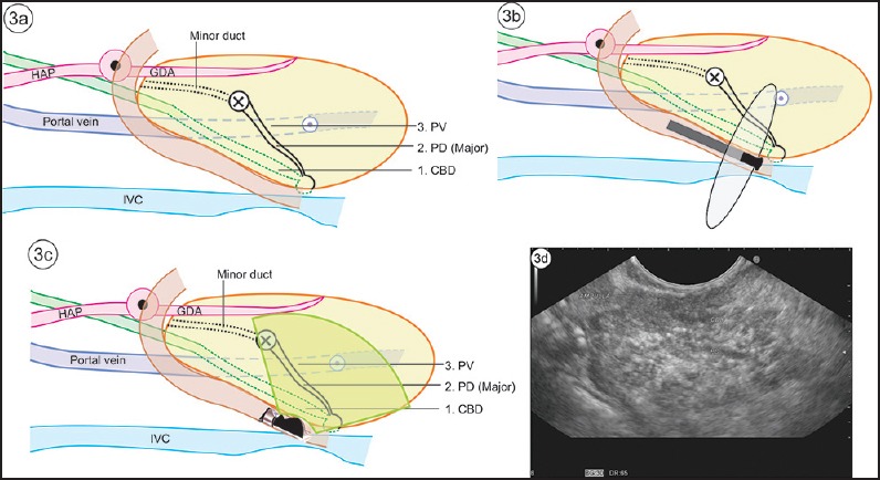
Figure 3.

(a) The course of CBD, major pancreatic duct and portal vein (PV), is shown in this figure. (b) The “stack sign” is a demonstration of the three structures in a stack by radial echoendoscope. (c) The stack is more often demonstrated by radial echoendoscope, but it is also possible to demonstrate the same stack with a linear echoendoscope. This has been termed “reverse stack sign” with a linear scope. It is easier to see the SMV continuing as portal vein. However, the axis oflinear imaging of the portal vein and SMV does not lie in the axis of CBD and pancreatic duct. (d) “Stack sign” of linear EUS
