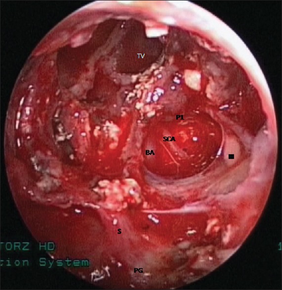
Figure 6.

Endoscopic view showing tumor cavity after complete removal of the tumor. TV = third ventricle, BA = basilar artery, P1 = left posterior cerebral artery, SCA = left superior cerebellar artery, III = left oculomotor nerve, S = pituitary stalk, and PG = pituitary gland
