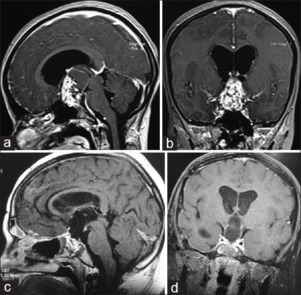
Figure 8.

Contrast-enhancing magnetic resonance imaging of a 14-year-old girl, sagittal (a) and coronal (b) views, showing a large solid, cystic, and calcified retrochiasmatic craniopharyngioma. Postoperative magnetic resonance sagittal (c) and coronal (d) imaging showing gross-total resection of the tumor
