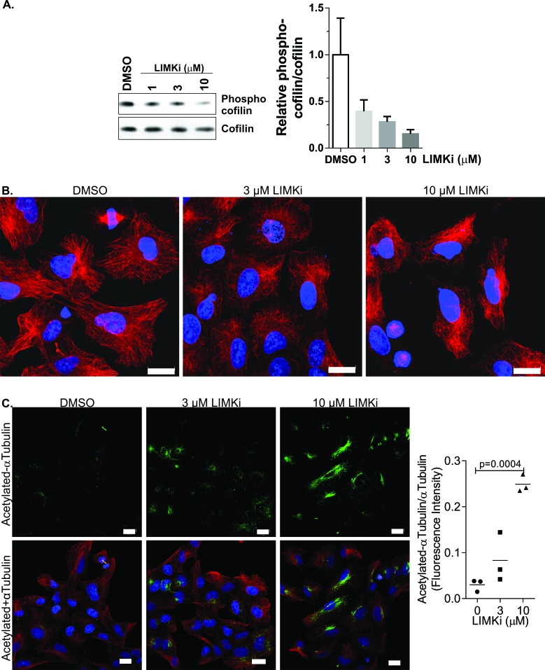
Figure 1. LIMK inhibition affects microtubule structures and acetylation.
A. A549 non-small cell lung adenocarcinoma cells were treated with LIMKi at the indicated concentrations for 24 hours, then cell lysates were Western blotted for phosphorylated and total cofilin. Graph indicates mean + SEM (n = 3). B. A549 non-small cell lung adenocarcinoma cells were treated as indicated for 24 hours, then fixed and stained with αTubulin antibody. Scale bar = 20 μm. C. A549 cells were treated as indicated for 24 hours, then fixed and stained with αTubulin (red) and acetylated αTubulin (green) antibodies. Nuclear DNA was stained with DAPI (blue). Scale bar = 20 μm. Immunofluorescence staining intensity of acetylated αTubulin was quantified with ImageJ software using a fixed intensity threshold, and normalized to total αTubulin immunofluorescence intensity levels. Statistical significance was analyzed by one-way ANOVA and Dunnett's post-hoc test (mean + SEM, n = 3).

