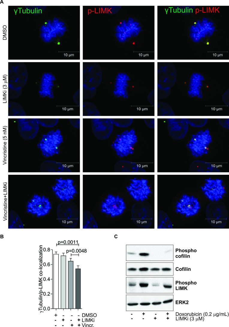
Figure 3. LIMK inhibition disrupts mitotic spindle integrity.
A. Co-localization of γTubulin (green) and phosphorylated LIMK (p-LIMK; red) was determined in A549 cells 24 hours after indicated treatments. Nuclear DNA was stained with DAPI (blue). B. Co-localization was analyzed on 10 randomly selected metaphase cells per indicated treatment. Pearson correlations of protein co-localization were quantified for each analyzed nucleus. Statistical significance of changes in protein co-localization were analyzed by one-way ANOVA and Tukey's multiple comparison post-hoc test (mean + SEM, n = 3). C. MCF-7 cells were treated with Adr (0.2 μg/ml) in the presence or absence of LIMKi (3 μM) for 24 h. Whole cell lysates were immunoblotted with antibodies against phospho-cofilin, cofilin, and phospho-LIMK. Equivalent protein loading was confirmed by ERK2 immunoblotting.

