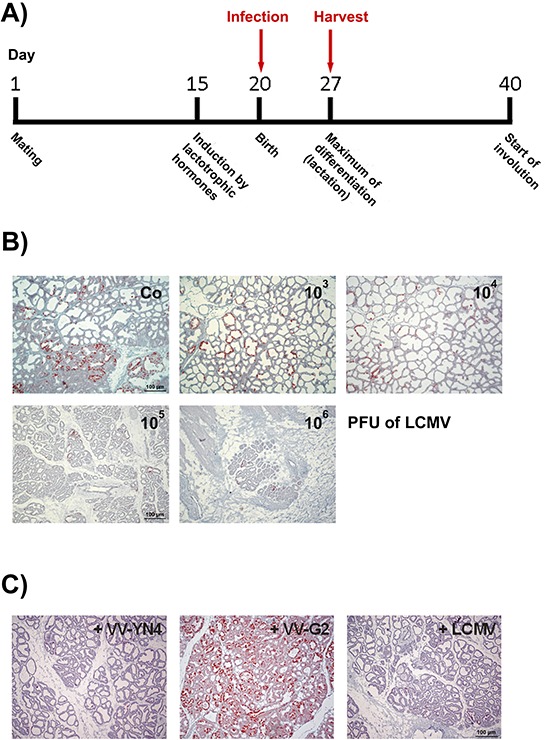
Figure 4. Infection of lactating NP8 mice with LCMV and VV recombinants for the analysis of a specific immune reaction against the LCMV NP118–126-epitope in T-AgNP.

A. Time line for harvesting the mammary glands after infection; four mice per group were infected with either different titers of LCMV or with 105 PFU of LCMV or VV recombinants – the relative amounts of T-Ag expressing cells are given as percentage in brackets. B. Immune-histological analysis for T-Ag expression of lactating mammary glands after infection with 103 [40–60%], 104 [10–20%], 105 [2–6%], or 106 PFU of LCMV [2–6%]; the preparation shown in the left upper corner presents an uninfected control tissue (Co) [100%]. C. Infection of transgenic mice with 105 PFU of VV recombinants containing either the NP (VV-YN4, left) [2–4%] or the glycoprotein-precursor of LCMV (VV-G2, middle) [~100%]. As control, NP8 mice were infected with LCMV (right) [2–6%].
