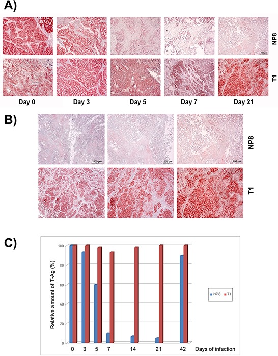
Figure 6. Specific elimination of T-AgNP expressing mammary tumor cells after LCMV infection.

A. NP8 mice (upper panels) or T1 mice (lower panels) bearing mammary tumors (diameter about 1.0 cm) were infected with 105 PFU of LCMV and tumors of mammary glands were prepared and stained for T-Ag at days 0, 3, 5, 7, 14 (not shown), 21, and 42 (not shown). B. Comparison of T-Ag expression (different magnifications) in tumors of NP8 (upper panels) and T1 mice (lower panels) 21 days after infection with LCMV. C. Demonstration of the transient elimination of tumor cells in NP8 mice (blue columns) and T1 mice (red columns) after virus infection by the loss of tumor-derived T-Ag (visual calculation of different fields within the stained samples).
