
Figure 1. The percentage of CD40+ (CD40+%) MDSC was significantly elevated in mouse spleens after tumor formation and was significantly higher in tumor tissue when compared with splenic tissue.
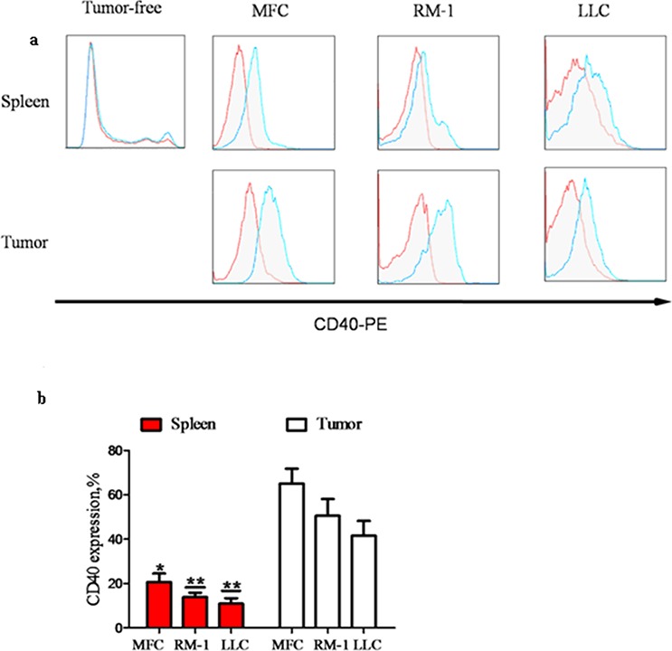
MDSC were isolated from the spleens of WT C57BL/6 mice without tumor inoculation (tumor-free; n = 5) or from the spleens and tumors of mice with tumors (diameter = 1 cm; n = 5) grown from subcutaneously injected MFC, RM-1 or LLC cells. CD40+% MDSC were analyzed using flow cytometry after staining isolated MDSC with PE-conjugated anti-CD40 antibody (CD40−PE; blue line) or PE-conjugated isotype-matched IgG control antibody (red line). a. Representative flow cytometry images showing minimal CD40+ cell detection in tumor-free mouse spleen, and a dramatic increase in CD40+% MDSC after tumor formation from all three different types of cancer cells. The highest CD40+% levels were detected in tumor tissues. b. CD40+% quantification in different groups of mice. *p < 0.05 and **p < 0.01, compared to the corresponding tumor tissues.