
Figure 1. 14-3-3ε induces HCC cell proliferation via β-catenin signaling activation.
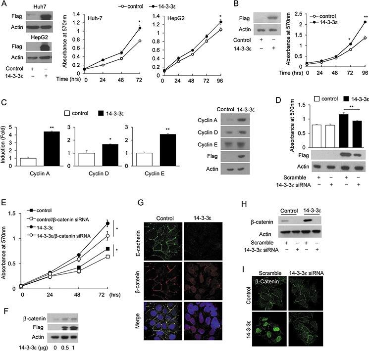
A. 14-3-3ε was transiently transfected in Huh-7 and HepG2 cells followed by an MTT assay (right panel). The expression of overexpressed 14-3-3ε was confirmed by Western blotting flag analysis (left panel). B. Cell proliferation was determined by an MTT assay in 14-3-3ε stably overexpressed (14-3-3ε) and control vector (control) Huh-7 cells (right panel). Expression of overexpressed 14-3-3ε was confirmed by Western blotting flag analysis (left panel). C. Cyclin A, cyclin D and cyclin E expressions were determined by RT-PCR (left panel) and Western blotting analysis (right panel) in control and 14-3-3 stable cells. (D, E) 14-3-3ε and β-catenin were knocked down by scramble control and siRNAs in control and 14-3-3ε stable cells for 48 hrs. Cell proliferation was determined by MTT analysis. Overexpressed 14-3-3ε expressions were determined by Western blotting flag analysis (left panel). F. Huh-7 cells were transfected with the indicated doses of 14-3-3ε overexpression vectors for 48 hrs. β-catenin and flag expressions were determined by Western blot analysis. G. E-cadherin and β-catenin expressions and subcellular localizations were examined by immunofluorescence confocal microscopy in control and 14-3-3ε stable cells. H. Control and 14-3-3ε stable cells were transfected with scramble and 14-3-3ε siRNA. β-catenin expression was determined by Western blot analysis. I. Control and 14-3-3ε stable cells were transfected with scramble and 14-3-3ε siRNA and subcellular localization of β-catenin was examined by immunofluorescence confocal microscopy. Actin was used as loading control for Western blotting analysis. Scale bars: mean ± SD. *, P < 0.05; **, P < 0.01.