
Figure 3. SDS-PAGE analyses of HQ5 and 800CW protein binding.
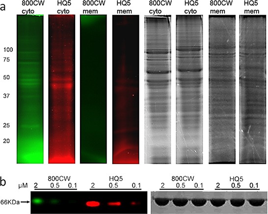
a. SDS-PAGE gel electropherogram of cytoplasmatic- and membrane fractions of 4T1-luc2 cell lysate, incubated with HQ5 or 800CW. Protein binding of HQ5 and 800CW was observed in the cytoplasmic but not in the membrane fraction. Coomassie blue staining confirmed the presence of proteins in both fractions. HQ5 and 800CW staining showed a different pattern, albeit with some common features. b. Binding of HQ5 or 800CW, at different concentrations (0.1, 0.5 and 2 μM), to bovine serum albumin (BSA).