
Figure 6. Monitoring anti-tumor efficacy in a EL4-CBG99-luc lymphoma mouse model of chemotherapy.
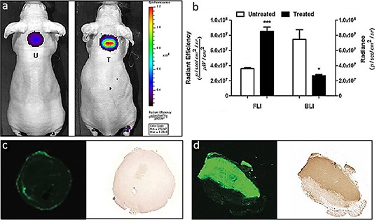
a. Representative whole body FLI images of tumor bearing mice treated with a combination of CTX and ETO and 24 hr later injected with 800CW. After another 24 h, in vivo whole body and ex vivo tumor FLI and BLI images were acquired and signal intensities were quantified. b. The BLI signals obtained from the treated animals were significantly lower as compared to those of the untreated controls (*p < 0.05). In contrast, the 800CW signals from the treated animals were significantly higher as compared to those of the untreated controls (***p < 0.001). c–d. Images of 800CW containing and a TUNEL stained tumor section of an (c) untreated tumor and a (d) treated tumor. The fluorescent signal obtained from a section of a treated tumor co-localized with TUNEL staining of the same section. Negligible 800CW fluorescence and TUNEL staining was observed in the untreated tumor. T = treated; U = Untreated.