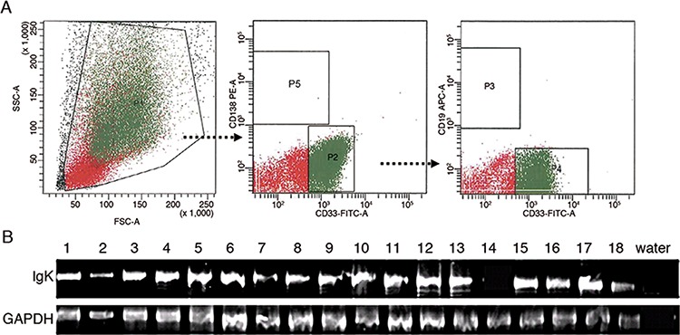
Figure 2. IGK expression in primary myeloblasts.

A. Flow cytometry sorting of myeloblasts. P1: mononuclear cells; P2: CD33+CD138− cells from P1; P3: CD19+ cells from P2; P4: CD33+CD19−cells from P2; P5: CD138+ plasma cells. The cells in P4 were defined as CD33+CD19−CD138− myeloblasts. The cells in P3 were used as a positive control. B. RT-PCR analysis detected IGK transcripts in myeloblasts from 17 of 18 AML patients.
