
Figure 6. Expression of apoptosis-associated genes in prostate cancer cells after treatment.
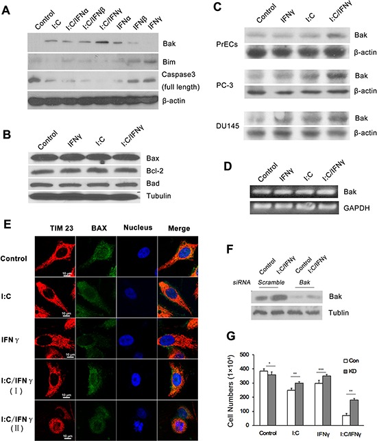
A. PC-3 cells were treated with 1,000 units/ml of individual IFN alone, or in combination with 1 μg/ml of poly I:C in the presence of lipofectamine. The expression of several apoptosis associated proteins was determined by Western blot analysis using antibodies to Bak, Bim and the full length of caspase 3. B. PC-3 cells were treated with 1 μg/ml of poly I:C, 1,000 units/ml of IFN γ alone, or a combination of both in the presence of lipofectamine for 14 hours. The expression of Bax, Bcl-2 and Bad was examined by Western blot analysis. C. PC-3, DU-145 and PrECs cells were treated as described above and the expression of Bak in the cells was examined by Western blot analysis. D. PC-3 cells were treated as described above. Total RNAs were isolated by using the Trizol Reagent (Invitrogen, Grand Island, NY) and the expression of Bak was determined by RT-PCR. The PCR primers for Bak are: sense 5′-CTG CCC TCT GCT TCT GAG GA-3′ and antisense 5′-CTGTCA GGA TGG GAC CAT TG-3′ E. Immunofluorescent staining of Bax in PC-3 cells after treatment. I:C/IFN γ I. pre-apoptotic cell; I:C/IFN γ (II): apoptotic cell. Tim 23: red (PE); Bax: green (FITC); and nucleus: blue (Hoechst). F. The expression of Bak in PC-3 cells was knocked down by through the utilization of a siRNA kit. G. Control and knock down cells were treated with 1 μg/ml of poly I:C, 1,000 units of IFN γ alone, or a combination of both in the presence of lipofectamine for 48 hours. The viable cells were analyzed by trypan blue exclusion assays and the cell numbers were averaged from three independent experiments. Error bars represent ± SEM, and Student's t test was used. *p < 0.05; **p < 0.01 and ***P < 0.001.