Figure 6.
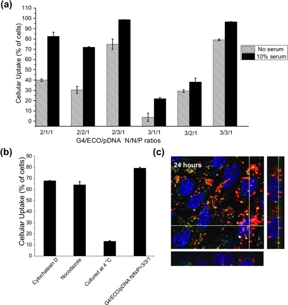
(a) Cellular uptake of G4/ECO/Cy3 pDNA nanoparticles cultured with ARPE-19 cells for 4 h. A range of N/P ratios were tested under serum-free and 10 % serum culture media. (b) Cellular uptake of G4/ECO/pDNA N/P ratio 3/3/1 particles cultured with ARPE-19 cells for 4 h in serum-free media under different inhibitory conditions (cytochalasin D (5 µg/mL), nocodazole (20 µM) and 4 C). (c) Comfocal fluorescence image of cytosolic delivery of G4/ECO/pDNA N/P ratio 3/3/1 particles. Late endosomes were stained with LysoTracker Green (green), nuclei were stained with Hoechst 33342 (blue) and DNA plasmid was labeled with Cy3 (red). At 24 h, nanoparticles mostly escape endosomal entrapment, shown by the red signals dispersed throughout the cytoplasm. Green signals represent late endosomes/lysosomes. Co-localization with late endosomes (yellow) appears minor.

