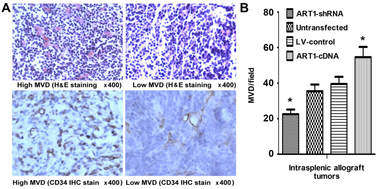
Figure 5.
Microvessel density (MVD) in mice intrasplenic allograft tumor tissues. (A) The expression of CD34 indicated 'hot spots' and low MVD area in intrasplenic allograft tumors (H&E and immunohistochemical staining; magnification ×400). (B) The comparison of MVD in intrasplenic transplanted argenine-specific adenosine diphosphate (ADP)-ribosyltransferase 1 (ART1)-cDNA CT26 tumors with transplanted ART1-shRNA CT26 cells, LV-control CT26 cells and untransfected CT26 cells. *P<0.05 vs. untransfected or LV-control CT26 cells; n=4 mice in each group.

