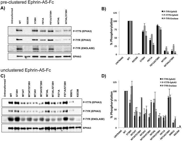
Fig 6. In vitro kinase assays probing for EphA3 autophosphorylation and enolase phosphorylation using full length EphA3 constructs in HEK293T cells.
A) HEK293T cells stimulated with pre-clustered Ephrin-A5-Fc. The EphA3 K653M mutant was used as a kinase-dead control. Phosphorylation of WT and mutant EphA3 was detected using phospho-tyrosine (phospho-EphA3 and phospho-enolase) and EphA3 activation loop (Y779) antibodies. B) Quantification of Western blot shown in (A). The histograms represent average values from 3 replicate experiments, with standard deviations shown as error bars. Values were normalized using WT as reference. C) Phosphorylation in un-clustered HEK293T cells. Phosphorylation of WT and mutant EphA3 was probed as described in A. D) Quantification of Western blots presented in (C). The histograms represent average values from 3 replicate experiments, with standard deviations shown as error bars. Values were normalized using WT as reference.

