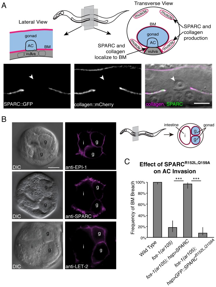
Fig 2. SPARC and type IV collagen colocalize in the gonadal BM and the collagen-binding pocket of SPARC is required to promote invasion.
(A) In the top panel, a schematic diagram shows the location of the BM, AC, and vulval cells in a lateral (left) and transverse (right) section of the worm. The transverse section also shows the body wall muscles, which are the predominant sites of type IV collagen and SPARC expression. Below, a lateral section shows SPARC::GFP (syIs115; left) and collagen::mCherry (center) fluorescence at the BM overlaid with a DIC image of an invaded AC (arrowhead; right). (B) Immunocytochemistry on fixed transverse worm sections shows that endogenous SPARC (anti-SPARC) and type IV collagen (anti-LET-2) localize with laminin (anti-EPI-1) in the BM. The schematic on the right indicates the approximate position of these sections and the location of the gonad (g) and intestine (i). All proteins colocalize in the gonadal BM. Type IV collagen and laminin localize uniformly in the gonadal BM, whereas SPARC appears to be more strongly enriched in the BM on the outer surface of the gonad. (C) The graph depicts the frequency of BM breach in animals overexpressing SPARC (hsp>SPARC) compared to animals overexpressing a form of SPARC (hsp>SPARCR152L,Q159A::GFP) that is unable to bind type IV collagen (n≥51 for each genotype). Error bars denote 95% confidence intervals with a continuity correction. *** denotes p<0.0005 by two-tailed Fisher’s exact test. Scale bars denote 5 μm.

