Fig 2.

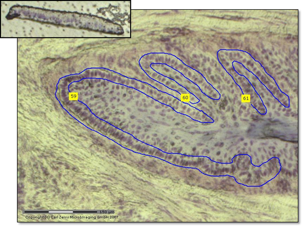
Selected section of lamellar basal epithelial cells (LBECs) stained with cresyl violet at 10X magnification from the PALM LCM microscope. Top insert demonstrates catapulted section of LBECs in adhesive cap.

Selected section of lamellar basal epithelial cells (LBECs) stained with cresyl violet at 10X magnification from the PALM LCM microscope. Top insert demonstrates catapulted section of LBECs in adhesive cap.